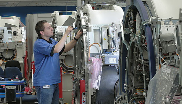

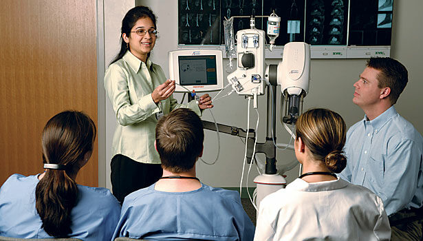
Advanced sensors, electronics and displays make these products difficult to design and assemble. The machines must capture images inside the body with low noise interference and convert them digitally so they can be displayed in real time on computer monitors and integrated into picture archiving and communication systems.
Traditionally, imaging and diagnostic machines are bulky, stationary devices that require patients to go to a special room in a hospital or doctor’s office. But, a new generation of compact devices comes directly to the patient.
For instance, during some recent college football bowl games, portable digital X-ray units played an important behind-the-scenes role. Sideline medical staff was also equipped with ultrasound technology that has typically only been available in hospitals. The portable equipment can deliver the same image quality of a stationary system in just seconds, allowing athletes to undergo much faster diagnosis.
Imaging and diagnostic equipment has made huge strides in the past decade. Cost has dropped dramatically, making devices more accessible to smaller medical practices and clinics. The size of the equipment has decreased, as well, making it much easier to use products in small spaces.
New technology has also led to innovative features that were unthinkable a few years ago. For instance, CT machines have evolved from a 16-slice scanner to a 256-slice scanner that can measure subtle changes in blood flow to detect the smallest blockages forming in blood vessels.
Radiology equipment featuring advanced image technology helps doctors make an accurate diagnosis. This includes being able to enhance the X-ray image by changing the color, size or contrast of the image or a specific part of the image.
“In addition, due to the general decrease in the size of equipment, radiographs and ultrasounds can now be developed directly in a small practice or clinic,” says Shalini Shahani Dewan, a medical industry analyst at BCC Research. “The technology has advanced to the point where images can be stored in an electronic database for future referencing purposes or even e-mailed to the patient directly.
“Due to their function and their interdependence, designing imaging and diagnostic equipment can be challenging,” adds Dewan, who says the U.S. medical imaging market is growing 9 percent annually. “Imaging equipment must be designed through visualizing the characteristics of the image which will be created. [On the other hand], diagnostic equipment must be designed through theorizing what characteristics of the image are required to provide an accurate diagnosis.”
During the recent Radiological Society of North America show in Chicago, medical device manufacturers displayed numerous innovations. For instance, GE Healthcare featured a new image-guided X-ray system that glides around like a robot.
The Discovery IGS 730 travels to the patient and backs away when needed, using a laser-guided positioning system. Its only tether is a sleeve of cables that trails the machine. The engineer who designed the new system got the idea by watching robotic warehousing equipment at a trade show.
Traditionally, there have been only two categories of imaging systems available. Physicians had to manipulate patients around a machine that was fixed to the floor or slide a large imaging system attached to rails and suspended from the ceiling.
The new robotic system knows precisely where it is because of a laser-guided indoor GPS system attached to the top of the device. The system constantly triangulates its position by sending and receiving laser beams to and from special reflectors attached to the wall.
A key component of the machine is a sleek, C-shaped arm attached to the mobile console. The arm slides automatically around the patient’s body.
“One end of the arm is capped with an X-ray source and the other with a detector,” says Hooman Hakami, president and CEO of GE Healthcare Interventional Systems. “The detector converts X-ray signals into real-time, high-quality images that allow the physician to precisely thread a medical device through the body to the target.”
Complex Designs
Medical imaging and diagnostic equipment is different than other types of medical devices. Unlike single-use, disposable products, such as syringes, tubing, anesthesia masks and intravenous catheters, MRI systems are durable machines that must be engineered for repeated use over a long period of time.
“The number of products made in each category is dramatically different—thousands or hundreds of thousands vs. millions and hundreds of millions,” says Len Czuba, president of Czuba Enterprises Inc., a leading medical device design firm. “This volume discrepancy accounts for differences in how the products are made, what materials are selected for use and how they are assembled.
“Cabinetry and housings must be made out of materials that can survive long-term use and repeated handling, cleaning, movement, and exposure to many different environments and chemicals,” says Czuba. “Because plastic is easier to process and easier to clean than metal, medical equipment manufacturers typically use thermoformed housings. Acrylics and polycarbonates are especially popular.”
Most diagnostic and imaging equipment is assembled with fasteners and adhesives. According to Robert Michaels, vice president of technical sales at Master Bond Inc., low-viscosity epoxies, silicones and encapsulation compounds are ideal for diagnostic equipment.
“These formulations feature high bond strength to similar and dissimilar substrates, offer high and low temperature resistance, withstand exposure to moisture and chemicals, and have long-term durability,” claims Michaels. “Specific grades are electrically conductive, thermally conductive USP Class VI approved, NASA low-outgassing certified and optically clear.”
Medical imaging and diagnostic equipment is getting smaller because of the continuous miniaturization of electronics, computerization and flat-screen technology. Engineers are using more embedded LEDs, dome arrays and silicone rubber keypad assemblies today, including touchscreen keypads that can be easily used even through surgical gloves.
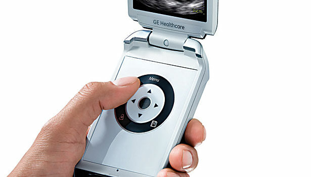
The latest generation of devices are lighter and easier to use than their predecessors. A good example is the Vscan, a pocket-sized, wireless ultrasound device that was developed by GE Healthcare engineers. The $7,500 device weighs less than 1 pound and replaces a laptop-sized, 20-pound device that costs $35,000. It can easily be taken from room to room and used in many different hospital, clinical and primary care settings.
Medical engineers are also focusing more efforts on designing ergonomically compatible features. “This means that the equipment must be easier to use, while instructions for use must be clearer and presented in an intuitive manner that helps avoid errors,” Czuba points out. “Design must also allow for easier and more reliable cleaning without cracks and crevices that can house ‘bugs,’ leading to cross-contamination from patient to patient rooms.
“Image acquisition is getting easier to read and interpret with more reliable data, since improvements in the computer hardware and software now allow for use of color and 3D reconstruction of the image target,” adds Czuba. “Medical professionals are demanding better graphics and colors, wireless telemetry and improved sensor technology.
“Improvements in every aspect of technology increases efficiency, functionality and reliability,” claims Czuba. “The [goal is to provide] better diagnosis and patient treatments. A 3D image of a broken or shattered bone, for instance, allows for a much better treatment.”
Electronic convergence allows engineers to incorporate more features that end users want, such as flexible devices that produce high-quality images at optimal speed. They’re incorporating membrane switches commonly used in the appliance industry and flat-screen displays widely found in iPhones, iPads and other popular consumer electronic devices.
“The challenge is to develop electronic components that can be upgraded to accommodate advancing technologies in a cost-effective manner,” says Bill Martineau, healthcare industry analyst at the Freedonia Group Inc. “[That’s especially true] in the area of touchscreen operation, optical fiber cables, miniaturization of cameras and electronic component interconnects.”
Fiber optics is increasingly replacing traditional copper cables for improved digital imaging and diagnostic purposes. According to Martineau, benefits of fiber optics include higher speeds and increased bandwidth, which improves the quality of the image by ensuring a stronger signal without distortion.
By providing more precise images, fiber optic cables allow for a shift from diagnostics to prevention. For instance, optical cables can remove ground loops and electromagnetic and radio frequency interference, allowing for a clearer video display on an MRI and X-ray image.
Future Trends
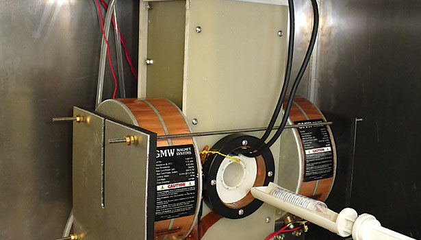
Evolving technology, such as molecular imaging and nanotechnology, is revolutionizing the diagnostic and imaging industry. For example, engineers at the University of California Berkeley have developed a new type of scanner that is different than traditional CT, MRI and nuclear imaging devices.
“The projection X-space magnetic particle imaging (MPI) scanner is the first of its kind,” claims Patrick Goodwill, a bioengineering student who developed the technology. “What makes the MPI technique unique is that it directly detects safe iron tracers with sensitivity far higher than what is possible in other medical imaging techniques such as MRI and CT. We anticipate that MPI will find use in rapid, safe angiography for assessment of cardiovascular health.
“MPI will save lives by giving doctors a way to assess cardiovascular health in real-time without the dangers of radiation and iodine inherent to the clinically dominant X-ray techniques,” adds Goodwill. “It will also give healthcare professionals a way to noninvasively study the body. MPI sees only an iron-oxide tracer and does not see tissue, [so it’s] comparable to nuclear imaging.”
One of the hottest trends in the medical device industry is molecular imaging. “The need to understand the fundamental molecular activities inside living organisms has led to increased interest in molecular imaging, which is a noninvasive procedure,” says Prasanna Vadhana Kannan, diagnostic imaging industry analyst at Frost & Sullivan Inc. “It enables the visualization of cellular functions without disturbing them.
“The enormous potential of this field is applicable to the diagnosis of diseases such as cancer, neurological and cardiovascular diseases,” Kannan points out. “The ability to image fine molecular changes opens up many possibilities for medical applications, such as early detection and treatment of disease. This has a huge economic impact due to the earlier and more accurate diagnosis.”
Molecular imaging differs from traditional imaging in that probes known as biomarkers are used to help image the targets or pathways. Biomarkers chemically interact with their surrounding tissue and in turn produce the image according to the molecular changes occurring within the area of interest.

